Figure 7.
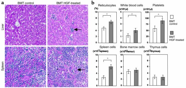
Augmentation of hematopoietic function by HGF in a syngeneic BMT model. Recipients were transplanted with 5 × 10⁶ bone marrow cells from syngeneic (BDF1) donors after 9 Gy of TBI. HGF-HVJ liposomes (or PBS) were injected on days 0 and 7. Ten days after BMT, histological examination was performed (a), and the peripheral blood cell profile, as well as the number of spleen cells, bone marrow cells, and thymus cells (b), was determined. (a) Hematoxylin and eosin staining of the liver and spleen in syngeneic BMT mice with or without HGF gene transduction. Liver tissue from HGF-treated GVHD mice showed hematopoietic foci containing granulocyte precursor cells and erythroblasts (arrow). Spleen tissue from HGF-treated GVHD mice showed marked extramedullary hematopoiesis along with numerous megakaryocytes (arrow). ×200. (b) The peripheral blood cell profile and the number of spleen cells, bone marrow cells, and thymus cells are shown as the mean ± SD of four mice. AP < 0.05.

