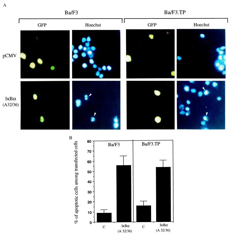
Figure 4.
Induction of apoptosis of Ba/F3 and Ba/F3.TP cells by the IκBα (A32/36) mutant. (A) Cells were transfected with 10 μg of the IκBα (A32/36) mutant or the same amount of the corresponding empty vector together with 5 μg of pEGFP plasmid. Ba/F3 and Ba/F3.TP cells were then cultured for 18 h in the presence or in the absence of IL-3, respectively, before evaluation of chromatin alteration by Hoechst staining. Arrows indicate apoptotic nuclei. (B) Quantification of apoptosis in control and IκBα (A32/36) transfected cells. Results are expressed as the percentage of apoptotic nuclei in transfected cells and represent the means ± SEM of three independent experiments. An average of 300 transfected cells were examined in each experiment.

