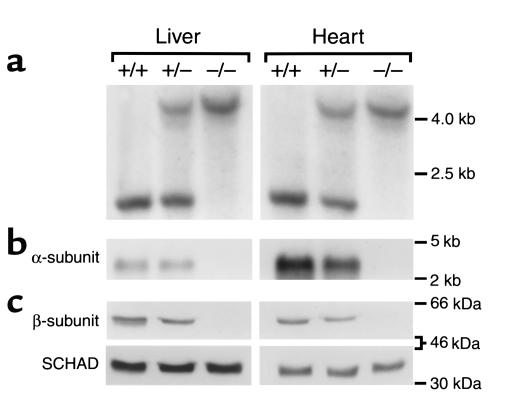
Figure 2.
Molecular characterization of MTP-deficient mice. Representative Southern blot (a), Northern blot (b), and Western blot (c) analyses using DNA, total RNA, and protein isolated from liver and heart tissues from pups sacrificed at birth with different genotypes: Mtpa+/+ (+/+), Mtpa+/– (+/–), and Mtpa–/– (–/–). Southern blot analysis was performed using EcoRI and NotI digestion. Mtpa+/+ mice had one band corresponding to the wild-type 2.2-kb fragment, Mtpa+/– mice had two bands corresponding to the wild-type 2.2-kb fragment and a mutant 4.1-kb fragment containing the neo cassette (see Figure 1), and Mtpa–/– mice had the mutant 4.1-kb band. An α-subunit cDNA probe was used in the Northern blot analysis, and antibodies raised against MTPβ or SCHAD were used in the Western blot analyses.

