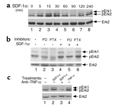
Figure 6.
SDF-1α activates Erk1/2 MAPK. (a) Kinetic analysis of SDF-1α–induced Erk1/2 MAPK phosphorylation. Whole-cell extracts were prepared after SDF-1α treatment of primary astrocytes for the indicated periods of time. Erk1/2 MAPK phosphorylation was then analyzed by anti–phospho-Erk1/2 Western blot (top panel). After stripping, the same membrane was blotted with anti-Erk2 antibodies (bottom panel). Data represent three experiments. (b) PD98059, but not PTX, suppresses SDF-1α–stimulated Erk1/2 MAPK phosphorylation. Murine primary astrocytes were first incubated with PD98059 for 1 hour or with PTX for 20 hours, and then stimulated with SDF-1α for 15 minutes. Whole-cell extracts were then prepared and subjected to anti–phospho-Erk1/2 Western blotting (top panel). After stripping, the same membrane was blotted with anti-Erk2 antibodies (bottom panel). Data represent three experiments. (c) TNF-α mediates SDF-1α–induced late Erk1/2 activation. Murine primary astrocytes were incubated with SDF-1α in the presence (lane 3) or absence (lane 2) of anti–TNF-α (5 μg/ml) for 2 hours or treated with TNF-α alone (lane 4) for 30 minutes. Whole-cell extracts were then prepared and subjected to anti–phospho-Erk1/2 Western blotting (top panel). After stripping, the same membrane was blotted with anti-Erk2 antibodies (bottom panel). Data represent three experiments.

