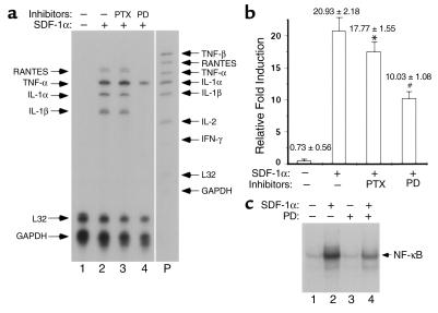
Figure 7.
SDF-1α–induced Erk1/2 activation is involved in TNF-α induction and NF-κB activation. (a) PD98059, but not PTX, suppresses SDF-1α–induced cytokine and chemokine mRNA expression. Murine primary astrocytes were incubated with medium alone, with PTX for 20 hours, or with PD98059 for 1 hour. Incubation was followed by SDF-1α stimulation for 2 hours. RNA was then prepared and analyzed by RPA. Unprotected probe bands are indicated with arrows on the right, and protected bands are shown with arrows on the left. Data represent four experiments. (b) PD98059, but not PTX, suppresses SDF-1α–induced TNF-α promoter reporter activity. Primary astrocytes in the presence or absence of PTX or PD98059 were transiently cotransfected with pGL3-mTNF (–1260) (0.5 μg) and pRL-TK (0.05 μg) constructs followed by SDF-1α stimulation for 6 hours. Cells were then lysed and subjected to luciferase activity assay. After normalization, mean ± SD relative luciferase activity was obtained from triplicate independent transfection (Student’s t test: *treatment with PTX + SDF-1α vs. SDF-1α alone, P = 0.1097; #treatment with PD + SDF-1α vs. SDF-1α alone, P = 0.0015). Data represent three independent experiments. (c) PD98059 suppresses SDF-1α–induced NF-κB activation. Murine primary astrocytes were treated with SDF-1α (lane 2) for 2 hours, with PD98059 for 3 hours (lane 3), or with PD98059 for 1 hour followed by SDF-1α for 2 hours (lane 4). Shown are results of EMSA with the IP-10 κB2 probes. Data represent three experiments.

