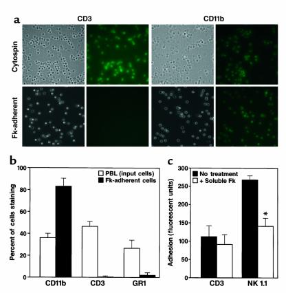
Figure 4.
Cell types adhering to Fk. (a) Peripheral blood leukocytes from CX3CR1+/+ mice were isolated as a buffy coat and either spun onto a glass slide (cytospin) or allowed to adhere to Ab- tethered Fk (Fk-adherent). Cells were then labeled with biotinylated Ab’s (CD3 or CD11b) and fluorescent streptavidin for identification. Bright-field and fluorescent images were captured. (b) Quantitation of cell types binding to Fk. Three fields were counted for each labeling condition, and the percentage of each cell type in the input population and Fk-adherent population is shown. The experiment shown is representative of two. (c) Adhesion of purified NK and T cells to Fk. NK (NK1.1+, CD3–) and T (CD3+, NK1.1–) cells were purified and fluorescently labeled. Cells were allowed to adhere to Ab-tethered Fk, in the presence or absence of soluble (100 nM) Fk. The experiment shown is representative of three. The asterisk indicates P < 0.005.

