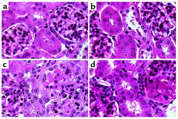
Figure 5.
Histology of control and nephritic kidneys. CX3CR1+/+ (a, c) and CX3CR1–/– (b, d) mice were preimmunized with normal sheep serum and then injected intravenously with normal sheep serum or nephrotoxic serum. Sections of kidneys harvested from mice immunized with normal sheep serum show normal renal morphology (a, b). In contrast, cortical sections obtained 21 days after injection with nephrotoxic serum (c, d) show proliferative and inflammatory glomerular changes, including hypercellularity, focal necrosis, increased matrix, thickening of capillary loops, and occlusion by matrix and microthrombi, as well as a periglomerular interstitial mononuclear cell infiltrate. (H&E-stained paraffin sections; original magnification, ×790). Shown are representative sections from 4 to 6 animals per group.

