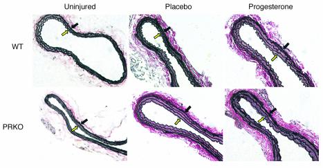
Figure 3.
Elastin-stained sections of uninjured and injured WT and PRKO mouse carotid arteries. Elastin-stained carotid artery sections representative of the mean medial area for each group are shown (×200). The media is delimited by the internal elastic lamina (open yellow arrow) and the external elastic lamina (black arrow).

