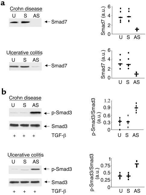
Figure 4.
Antisense to Smad7 restores TGF-β1 signaling in both CD and UC LPMCs. (a) Treatment of CD and UC LPMCs with a specific Smad7 antisense but not a sense oligonucleotide inhibits Smad7 expression. CD and UC LPMCs were cultured in the absence (U, unstimulated) or presence of a specific Smad7 antisense (AS) or control sense (S) oligonucleotide for 24 hours. Arrows indicate Smad7 detected by a specific polyclonal antibody. The example is representative of three separate experiments analyzing in total LPMCs from five CD or five UC patients. Quantitative data are shown in the right panel as measured by densitometry scanning of Western blots. Values are expressed in arbitrary units (a.u.). Each point represents the value (a.u.) of Smad7 in LPMCs taken from a single subject. Horizontal bars indicate the median value. (b) Inhibition of Smad7 restores the TGF-β1–induced Smad3 phosphorylation in both CD and UC LPMCs. LPMCs were cultured in the absence (U, unstimulated) or presence of a specific Smad7 antisense (AS) or control sense (S) oligonucleotide, and then stimulated with 1 ng/ml TGF-β1 for 1 hour. The right panel shows quantitative analysis of active/inactive Smad3 ratio in LPMC from five patients with CD and five with UC, as measured by densitometry scanning of Western blots. Values are expressed in arbitrary units (a.u.). Each point represents the value (a.u.) of active/inactive Smad3 ratio in LPMCs taken from a single subject. Horizontal bars indicate the mean.

