
Figure 3.
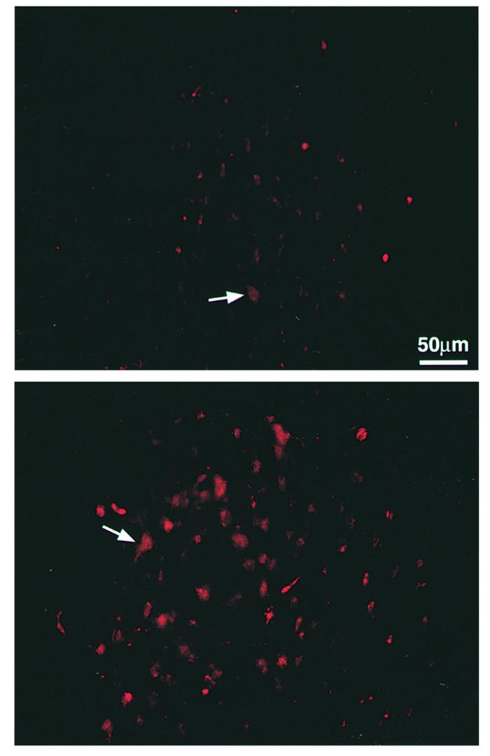
Retrograde labeling of cell bodies in the red nucleus. Spinal contusion was preceded 7 days earlier by immunization with spinal cord homogenate emulsified in CFA (containing 0.5 mg/ml bacteria) or by injection with PBS in the same adjuvant (Figure 2a). Three months later, two rats from each group were reanesthetized and the dye rhodamine dextran amine (Fluoro-ruby) was applied below the site of contusion. Five days later, the rats were sacrificed and their brains were excised, processed, and cryosectioned. Sections taken through the red nucleus were inspected and analyzed qualitatively and quantitatively by fluorescence and confocal microscopy. Significantly more labeled rubrospinal neurons were seen in slices from the immunized rats (bottom) (BBB score = 8) than from the PBS-treated rats (top) (BBB score = 5.5).