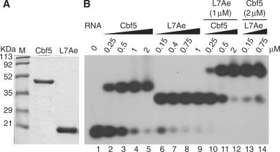
Figure 2.
Reconstitution of Cbf5–Pf9, L7Ae–Pf9 and Cbf5–L7Ae–Pf9 sub-complexes. (A) Coomassie blue staining of purified recombinant Cbf5 and L7Ae following PAGE. M lane contains protein standards. (B) Gel mobility shift analysis of 5′-end labelled Pf9 RNA, alone (lane 1) or with increasing amounts of Cbf5 and/or L7Ae as indicated. Complexes were separated on an 8% native gel and visualized by autoradiography.

