Figure 5.
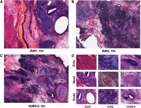
Teratoma formation SCID mice studies. The hESC lines ZUN1 (A), ZUN2 (B) and the parental line HUES8 (C) were injected into the hind leg of male SCID mice, and the teratoma formation was followed by a simple grading system (see Materials and Methods section). When teratomas were full developed (1–2 g tissue weight, 6–8 weeks), they were harvested and processed by routine histological procedures, followed by heamatoxilin/eosin staining. (D) Detailed histology of ectodermal (epithelium, neuroectoderm), mesodermal (bone in red, cartilage in blue) and endodermal (glandular structures) structures are shown for each hESC line.

