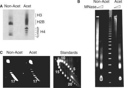
Figure 1.
Assembly of acetylated and non-acetylated chromatin. (A) Triton acid urea gel showing acetylated (Acet) and non-acetylated (Non-Acet) histones purified from CV1 cells. Numbers indicate the lysines modified. (B) Micrococcal nuclease ladders of the assembled non-acetylated (Non-Acet) and acetylated (Acet) arrays. The marker is a 123 bp ladder. (C) Supercoiling analysis of acetylated (Acet) and non-acetylated (Non-Acet) templates. The standards show supercoiling analysis of templates with known nucleosome densities.

