Figure 2.
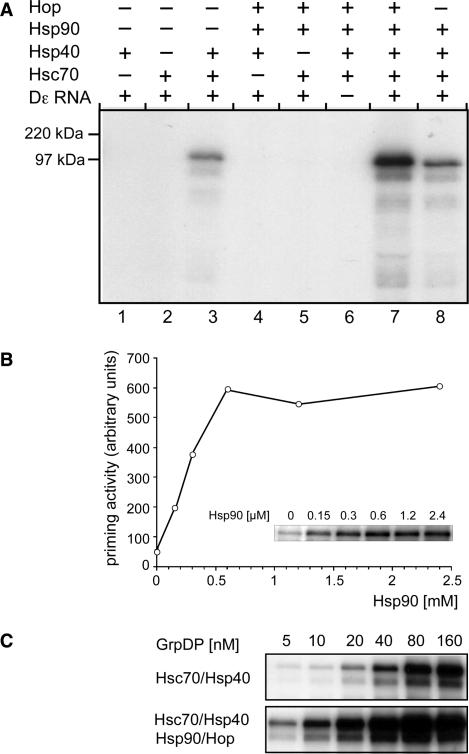
Chaperone dependence of priming-active P–Dε complex formation. (A) Stimulatory role of Hsp90. P protein–Dε complexes were reconstituted from GrpDP (150 nM) in the presence of the indicated chaperones and subsequently subjected to priming assays. Samples were separated by SDS–PAGE and the 32P-labeled P protein was visualized by autoradiography. (B) Effect of Hsp90 concentration on complex reconstitution. P protein–Dε complexes were reconstituted from GrpDP (10 nM), and fixed concentrations of Hsc70, Hsp40, Hop and Dε RNA plus varying concentrations of Hsp90 as indicated. Active complexes were detected by priming assays. 32P-labeled P protein was visualized by phosphorimaging (inset), and the quantified values were plotted against the Hsp90 concentration. (C) Effect of P protein concentration on complex reconstitution. P–Dε complexes were reconstituted with increasing concentrations of GrpDP in the presence of fixed concentrations of Hsc70 and Hsp40 only (upper panel), or in the additional presence of Hsp90 and Hop (lower panel). Complex formation was analyzed by priming assays as in (A).

