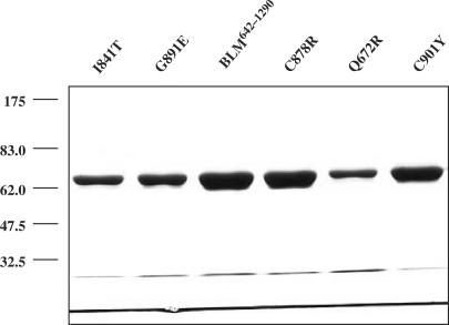
Figure 4.
SDS-PAGE analysis of the purified BLM642–1290 and various mutants. The proteins (indicated above the figure) were resolved in 10% SDS-polyacrylamide gel and stained with Coomassie blue. The amounts of the proteins used were 3.5 µg for BLM, C878R and C901Y and 1.5 µg for I841T, G891E and Q672R. The positions of marker proteins (in kDa) are indicated on the left.

