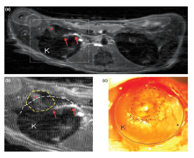
Figure 2. Representative imaging of vascularization of iron-labeled islet grafts on day 14.

(a) Postcontrast magnetic resonance imaging (MRI) at day 14 shows a contrast-enhancement (red arrows) in the vicinity of labeled transplanted islets. Similar densities were not observed in the contralateral kidney. (b) Expanded view of ‘A’ revealing selective enhancement (asterisk) within the islets (dotted line) and the linear density (possibly a communicating vessel) (arrows). (c) Following MRI, the kidney (circle) was exposed in vivo and a low power photomicrograph was taken of the transplanted islets (dotted line). Note the blood vessels on the kidney surface near the transplant (a, b, and c are from the same kidney).
