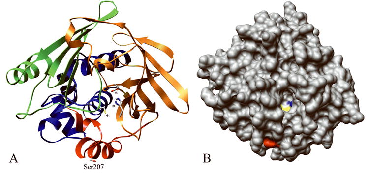
Figure 5.

Computational structures of rNat3 1 and rNat3 2 modeled after human NAT1 crystal structure (PDB ID# 2IJA). A The location of residue Ser207 (labeled) in the inter-domain region (red), and catalytic triad residues Cys68, His107, Asp122 are demonstrated (marked by asterisks). Protein domains I, II, and III are indicated by ribbon colors blue, orange, and green, respectively. B The rNat3 2 homology model with protein surface shown, demonstrates the location of Ser207 on the protein surface (red), near the active site opening in which the catalytic Cys68 (yellow) and His107 (blue) are visible.
