
Figure 2. A representative electron micrograph illustrating the relative distribution of immunogold labeling of GLUT1 glucose transporters in endothelial cells and astrocytic endfeet.
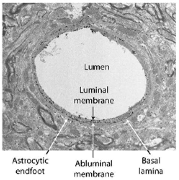
GLUT1 glucose transporters were detected by a combination of an antibody raised against purified human erythrocyte GLUT1 (Wheeler et al. 1982) and Alexa Fluronanogold Fab fragment goat anti rabbit Nanoprobe. Arrows, endothelial GLUT1; arrowheads, astrocytic GLUT1.