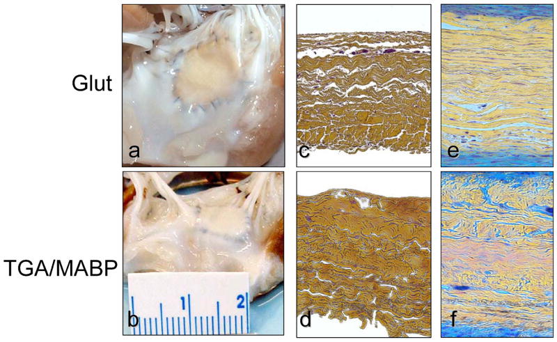
Figure 3.
Explant morphology comparing the following: (a) A gross specimen photographs of the glutaraldehyde valvuloplasty site; (b) The TGA valvuloplasty site with more exuberant overgrowth of connective tissue compared to glutaraldehyde fixation (see 3a); Cross-sections (c–f) of: (c) Unimplanted glutaraldehyde fixed bovine pericardium; (d) Unimplanted TGA-MABP pretreated bovine pericardium; (e) A 4 week explant specimen demonstrating overgrowth of the glutaraldehyde fixed specimen with host tissue with intense (blue) glycosaminoglycan staining; (f) A 4 week TGA-MABP explant demonstrating qualitatively greater overgrowth than observed with glutaraldehyde fixation (see 3f). Movats stain for figures c–f, original magnification 50x.

