
Figure 11.
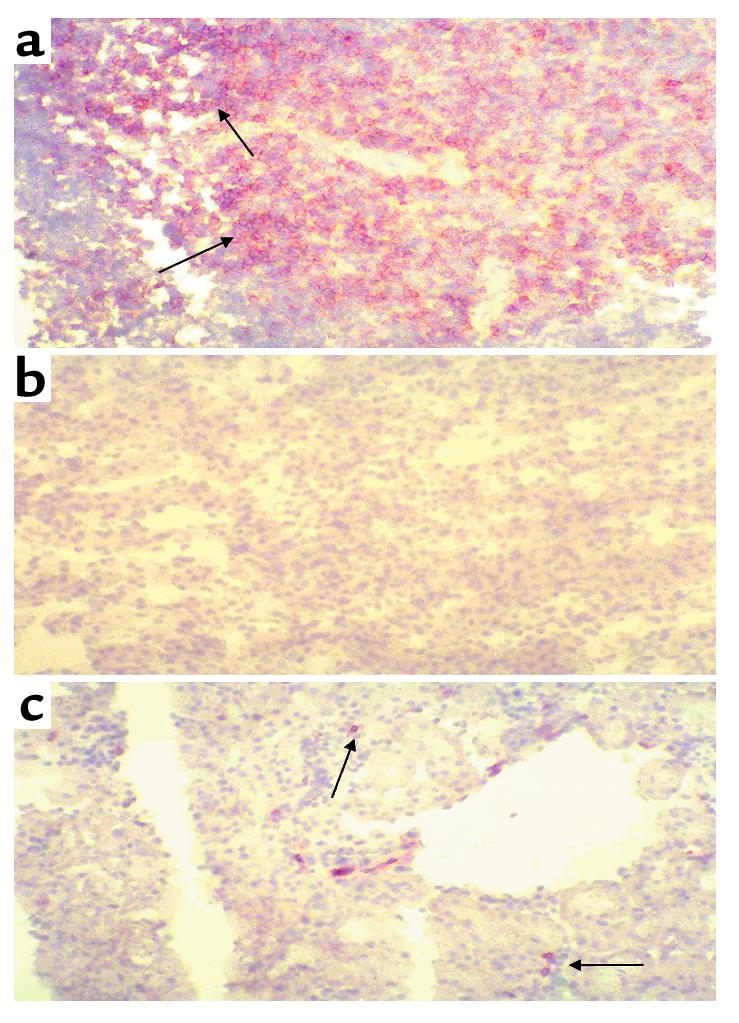
CD4+ T cells infiltrating into the postischemic kidney. (a) Positively stained wild-type spleen showing the presence of CD4+ T cells. This is represented by brown rings surrounding positive cells. (b) Wild-type (no IRI) kidney stained with an antibody for CD4. (c) A 24-hour postischemic kidney obtained from a wild-type control mouse, showing a small infiltration of CD4+ T cells. ×50.