
Figure 6. Sply05091 egg chambers exhibit pronounced nuclear and actin degeneration.
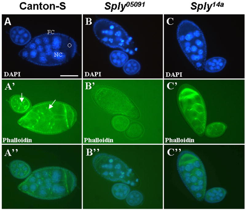
Dissected ovarioles containing egg chambers from 4-day old female flies co-stained with DAPI and phalloidin for detection of the nuclei and actin structures, respectively. (A–C) DAPI staining reveals increased membrane blebbing and nuclear condensation of Sply05091 mutant nurse cells (NC), oocyte (O) and enveloping follicle cells (FC) and minimal cell death of Canton-S and Sply14a revertant egg chambers. (A′–C′) Phalloidin staining shows loss of actin filaments such as the ring canals (arrow). (A″–C″) Overlay of the co-stained images. Bar, 50 μm.