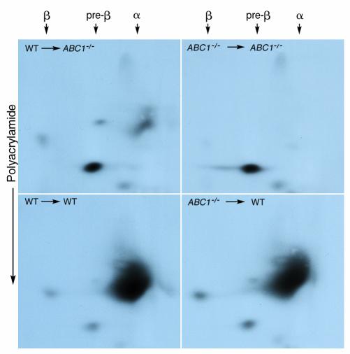
Figure 3.
Distribution of mouse apoAI by two-dimensional gel electrophoresis. Fourteen weeks after transplantation, 15 μl of pooled plasma from each transplant group was applied to electrophoreses in 0.75% agarose gel, which was then applied to a 3–16% polyacrylamide gradient gel. Plasma proteins were transferred to nitrocellulose membranes, and mouse apoAI was detected using a rabbit polyclonal Ab.

