
Figure 1. Schematics of the anthrax toxin-Soc fusion recombinants.
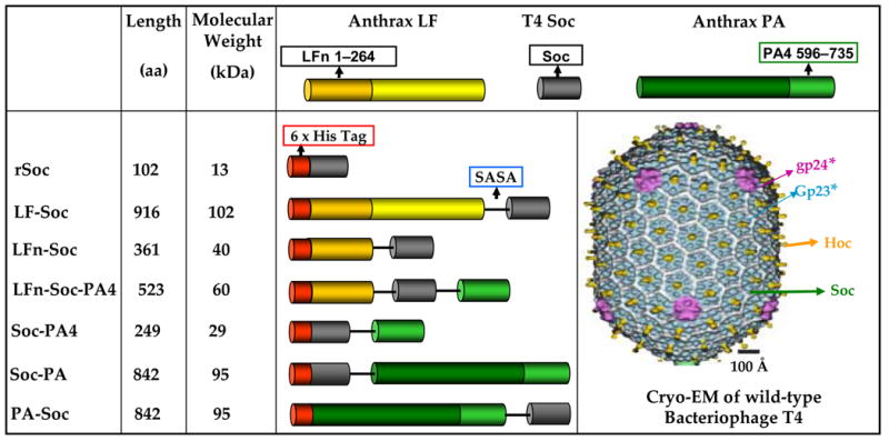
The anthrax toxin genes, PA, PA4, LFE687C, and LFn, were fused to the N- and/or C- terminus of Soc via the linker, SASA, as shown. Hexa-histidine tag was fused to the N-terminus of each gene fusion. The fusions were constructed using the PCR-based splicing by overlap extension (SOE) strategy. The numbers in the boxes represent the aa numbers of the respective proteins. The cryo-EM reconstruction of phage T4 capsid1 is shown to indicate the disposition of the capsid proteins; gp23* subunits are shown in blue, gp24* in purple, Hoc in yellow, and Soc in white.