
Figure 8. Bipartite display using both Soc and Hoc.
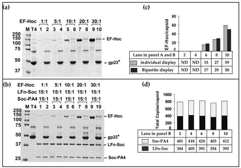
In vitro binding and analyses were performed as described in Materials and Methods and legend to Figure 3. About 2 × 10¹⁰ hoc−soc− T4 phage were incubated with EF-Hoc (a) or a mixture of EF-Hoc, LFn-Soc, and Soc-PA4, at the ratios indicated (b) (a): display of EF-Hoc. (b): bipartite display of EF-Hoc, LFn-Soc, and Soc-PA4. Lanes: M, mol. wt. standards; T4: hoc− soc− phage control; 1, 3, 5, 7, and 9: unbound proteins; lanes 2, 4, 6, 8, and 10: phage-bound proteins. (c) and (d): Histograms depicting the copy number of displayed proteins at different ratios, as calculated from the respective lanes in panels (a) and (b).