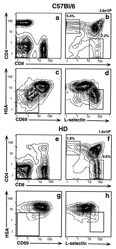
Figure 5.
The HD defect maps to the hematopoietic compartment. Sublethally irradiated RAG2−/− recipients were reconstituted with T cell-depleted bone marrow from wt (a–d) or HD (e–h) mice. PBLs and thymocytes were collected after 4 wk and stained with antibodies against CD4, CD8, HSA, CD69, and l-selectin. Staining profiles are shown for total PBLs (a and e), total thymocytes (b and f), and SP CD4+ thymocytes (c, d, g, and h). Insets in c, d, g, and h indicate the position of the most mature SP CD4+ subpopulation which is present in wt but not in HD mice.

