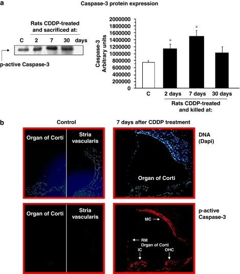
Figure 3.
Active caspase-3 expression. (a) A representative western blot of active p-17 caspase-3 protein expression levels in whole cochlea extracts of rats killed at 2, 7 or 30 days after CDDP injection is shown (left). Densitometric analyses of previous western blots are also shown (right). Results are expressed as arbitrary units; *P⩽0.05 vs control. (b) Immunostaining of active caspase-3 in the nuclei of inner ear cells from rats 7 days after a single dose of cisplatin (5 mg kg−1). Cryosections from the cochleae of exposed animals were stained (see Methods section). Increased nuclear caspase-3 immunostaining is seen in several cell populations after administration of cisplatin. MC, marginal cells of stria vascularis; RM, Reissner's membrane; OHCs, outer hair cells; IC, interdental cells of the spiral limbus. Magnification × 400. CDDP, cis-diamminedichloroplatinum II.

