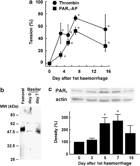
Figure 5.
Time course of enhancement of contractile responses and changes in PAR1 expression in the basilar artery without the endothelium. (a) The changes in the maximal tension developed by 1 U ml−1 thrombin and 10 μM PAR1-AP over 15 days after the initial injection of autologous blood. *P<0.05 vs day 0. (b), Representative immunoblot of PAR1 in rabbit femoral artery obtained 1 week after balloon injury as reported (Fukunaga et al., 2006) and in rabbit basilar artery obtained on days 0 and 7 after subarachnoid haemorrhage (SAH). The molecular weight is indicated on the left. (c) Immunoblot analysis of the change in the expression of PAR1 protein in the basilar artery after SAH. The level of expression seen on day 0 was assigned to be 100%. The data are the mean±s.e.m. (n=4–5 for (a), n=3 for (c), *P<0.05 vs day 0.

