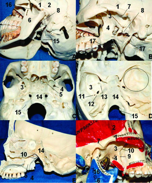
Figure 1.
Limits of the ITF and bone relationships. (A) Oblique lateral view of the ITF. (B) Same view after removal of the mandible. (C) Inferior aspect of the cranium. (D) Interior view of the cranial base. The circle on the right middle fossa represents approximately the correspondence of the ITF in the middle fossa. (E) Lateral view of the ITF after sagittal paramedian section. (F) Note that the depression of the mandible (open mouth) gives more access to the ITF laterally. 1, zygomatic process of the temporal bone; 2, temporal fossa; 3, greater wing of the sphenoid; 4, lateral pterygoid plate; 5, medial pterygoid plate; 6, mandibular ramus; 7, articular tubercle of the temporal bone; 8, spine of the sphenoid bone; 9, pterygomaxillary fissure; 10, pterygopalatine fossa; 11, foramen spinosum; 12, foramen ovale; 13, foramen rotundum; 14, clivus; 15, occipital condyle; 16, coronoid process; 17, styloid process. ITF, infratemporal fossa.

