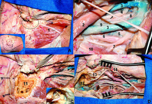
Figure 10.
Combined infratemporal and posterior fossa approach. (A) After the incision (inset) the flap is reflected anteriorly and the external meatus is sectioned and closed in a blind sac as described by Ugo Fisch (oval). (B) The neurovascular structures are exposed in the neck to proximal control. The arrow indicates the greater auricular nerve. (C) The petrosectomy is performed. The inset shows the facial nerve, the ossicles, and semicircular canals. (D) Final anatomical view of the neurovascular structures in the neck and the presigmoid and temporal fossa dura after petrosectomy. The semicircular canals are preserved and the facial nerve is transposed anteriorly. 1, great auricular nerve; 2, digastric muscle (posterior belly); 3, duplicated internal jugular vein; 4, glossopharyngeal nerve; 5, internal carotid artery; 6, sympathetic trunk; 7, accessory nerve; 8, hypoglossal nerve; 9, vagus nerve; 10, inferior oblique nerve; 11, mastoid drilled; 12, facial nerve; 13, semicircular canals; 14, superficial temporal artery; 15, transverse process of C1.

