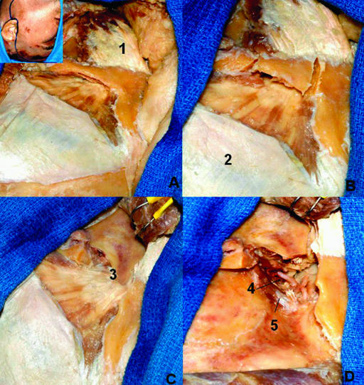
Figure 12.
Zygomatic approach. Part of the deep temporal fascia was removed to show the muscular fibers. (A) Preauricular incision and anterior displacement of the flap. (B) Section of the zygomatic arch. (C) The masseter and the zygomatic arch are displaced inferiorly. (D) The coronoid process is sectioned and displaced upward with the temporal muscle. 1, masseter muscle; 2, deep temporal fascia; 3, coronoid process; 4, maxillary artery; 5, lateral pterygoid muscle (upper head).

