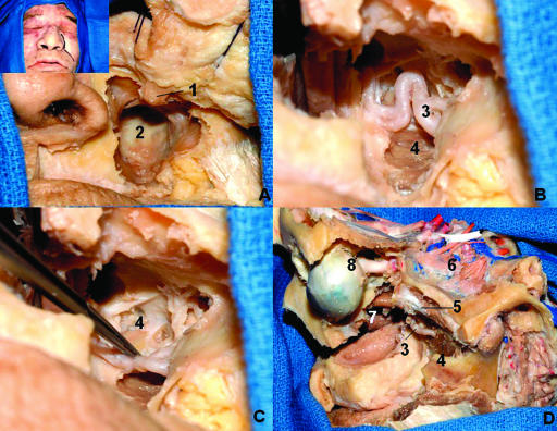
Figure 13.
Lateral transantral maxillotomy. (A) The anterior and lateral walls of the maxilla are resected. (B) After removal of the sinus mucosa, the posterior wall is drilled out, exposing the ITF. (C) The maxillary is displaced to show the lateral pterygoid plate. (D) This dissection exposes the ITF via an anterior and lateral view. The maxilla was totally removed. 1, infraorbital nerve; 2, posterior wall of the maxilla; 3, maxillary artery; 4, lateral pterygoid plate; 5, lateral pterygoid muscle (upper head); 6, lateral pterygoid muscle (lower head); 7, nasal cavity; 8, eyeball and optic nerve. ITF, inferior temporal fossa.

