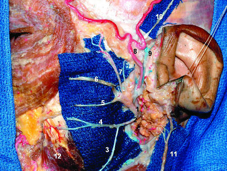
Figure 3.
Lateral view of the superficial dissection of the left face. The majority of the parotid tissue was removed to expose the facial nerve branches. A blue field was put under the nerves to enhance them. The distal ramification as well as the parotid plexus are not shown. 1, facial nerve (superior trunk); 2, facial nerve (inferior trunk); 3, cervical branch; 4, mandibular branch; 5, buccal branch; 6, zygomatic branch; 7, temporal branches; 8, superficial temporal artery; 9, superficial temporal vein; 10, auriculotemporal nerve; 11, great auricular nerve; 12, masseter muscle.

