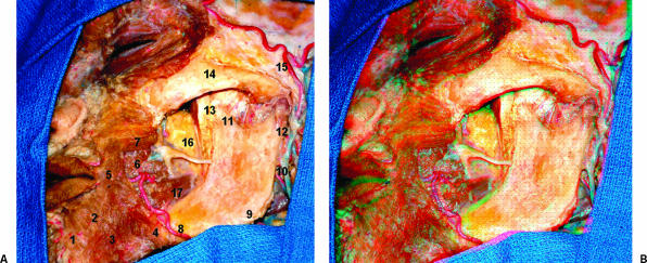
Figure 4.
(A) Lateral view of the deep dissection of the left face. The masseter muscle was removed. Note the insertion of the temporal tendon into the coronoid process and anterior aspect of the mandible. 1, mentalis muscle; 2, depressor labii inferioris; 3, depressor anguli oris; 4, fibers of the platisma muscle; 5, orbicularis oris muscle; 6, risorius muscle; 7, zygomatic major muscle; 8, facial artery; 9, angle of the mandible; 10, retromandibular vein; 11, coronoid process; 12, condylar process; 13, coronoid process; 14, zygomatic bone; 15, superficial temporal artery; 16, buccal fat pad; 17, medial pterygoid muscle. (B) The figure shows the stereoscopic view.

