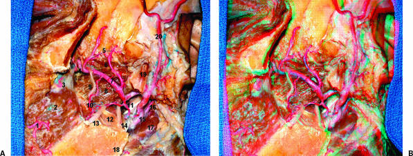
Figure 5.
(A) Lateral view of the ITF. The zygoma and part of the mandible were removed. The condylar process was left to show the insertion of the lateral pterygoid muscle in the pterygoid fovea in the neck of the mandible. 1, facial artery; 2, buccinator muscle; 3, posterior superior alveolar artery; 4, sphenopalatine artery; 5, anterior and posterior deep temporal arteries; 6, lateral pterygoid muscle (upper head); 7, maxillary artery; 8, lateral pterygoid muscle (lower head); 9, buccal nerve; 10, buccal artery; 11, styloid process; 12, medial pterygoid muscle; 13, lingual nerve; 14, inferior alveolar nerve; 15, external carotid artery; 16, posterior auricular artery; 17, digastric muscle (posterior belly); 18, angle of the mandible; 19, condylar process; 20, superficial temporal artery.(B) The figure shows the stereoscopic view. ITF, infratemporal fossa.

