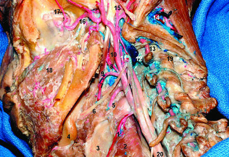
Figure 8.
Lateral view of the ITF and neck, emphasizing the branches of the external carotid artery. 1, superior thyroid artery; 2, inferior pharyngeal constrictor muscle; 3, thyroid cartilage; 4, submandibular gland; 5, lingual artery; 6, hypoglossal nerve; 7, facial artery; 8, ascending palatine artery; 9, stylohyoid muscle; 10, styloglossus muscle; 11, ascending pharyngeal artery; 12, internal jugular vein sectioned; 13, vertebral artery; 14, suboccipital triangle with venous plexus and muscular branches from the vertebral artery; 15, styloid process and posterior auricular artery; 16, maxillary artery; 17, infraorbital nerve; 18, buccinator muscle. ITF, infratemporal fossa.

