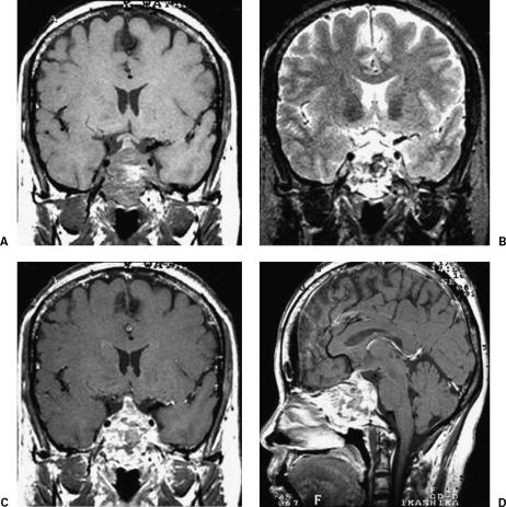
Figure 1.
Magnetic resonance images of the clival tumor before the initial operation. (A) T1-weighted, (B) T2-weighted, and (C,D) T1-weighted images with contrast medium. The tumor shows high-signal intensity on the T1-weighted image and low-signal intensity on the T2-weighted image, probably due to intratumoral hemorrhage. The tumor was enhanced with gadolinium-DTPA.

