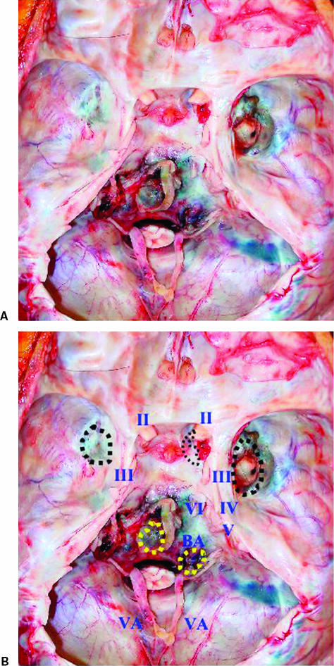
Figure 4.
(A,B) Photographs obtained at autopsy showing the internal skull base. The dura mater is uniformly intact and tumor tissue adheres to four locations extradurally (dotted circle): the bilateral middle fossae and the mid and lower portions of the clivus. The pituitary fossa and the posterior clinoid process show thinning and the pituitary gland cannot be visualized. The tumor around the lower clivus compresses the lower cranial nerves bilaterally.

