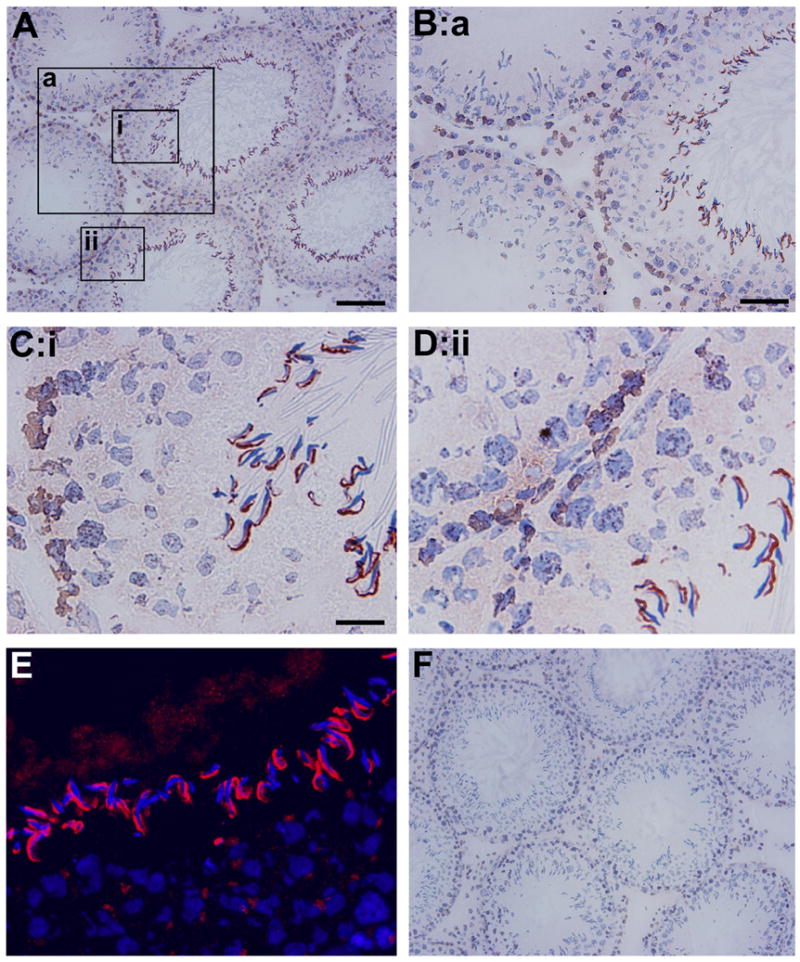
Fig. 2. Localization of CAR in the seminiferous epithelium of adult rat testes by immunohistochemistry.

A: Frozen sections of adult rat testes were stained using a rabbit anti- CAR polyclonal antibody. Signal was detected at the basal compartment of the epithelium in virtually all stages of the epithelial cycle. However, strongest staining was found at the apical ectoplasmic specialization in stage VIII tubules. B: Magnified view of the boxed area “a” in A, showing CAR staining at the basal compartment, which is consistent with its localization at the blood-testis barrier. C-D: Corresponding to boxed area (i) and (ii). Sickle shaped CAR staining was concentrated at site of apical ES in stage VIII seminiferous tubules, where elongate spermatids anchor onto Sertoli cells in the epithelium. CAR staining was also found at the site of blood-testis- barrier. E: Localization of CAR by immunofluorescent staining. Sections were incubated with a rabbit anti-CAR, to be followed by a donkey anti-rabbit IgG-Cy3 conjugate. Cell nuclei were visualized by DAPI staining. Merged image of CAR and DAPI staining identified CAR at the apical and round spermatids. F: Control experiment in which testis sections were stained with normal rabbit IgG at the same dilution as the primary antibody shown in A-E. Scale bar = 100 μm in A, which also applies to F Scale bar = 50 μm in B Scale bar = 15 μm in C, which also applies to D & E.
