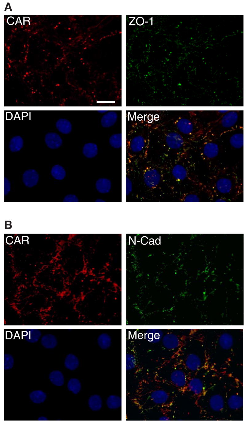
Fig. 4. CAR localize at cell-cell contacts of Sertoli cells.
Sertoli cells were cultured at low density (1 × 10⁵ cells/cm2) for 3 days before staining. Areas of co-localization appear as orange. Immunofluorescent micrograph demonstrates that CAR was heavily concentrated at inter-Sertoli tight junctions, though staining occasionally was also seen close to the nucleus. A: Cells were incubated with a rabbit anti-CAR polyclonal IgG as primary antibody, followed by a goat-anti- rabbit CY3 conjugated secondary antibody. A mouse anti-ZO-1 FITC conjugate was used to locate inter-Sertoli tight junctions. B: Cells were incubated with a rabbit anti-CAR (H-300) polyclonal antibody, along with a mouse anti-N-cadherin monoclonal antibody. N-cadherin is a known component of the inter-Sertoli cell junctions at the blood-testis barrier. Scale bar = 20 μm in Fig. 4A, which also applies to Fig 4B. This experiment was repeated at least 4 times over a period of 18 months using different batches of Sertoli cells where cultures were terminated on either day 2 (n =2) or day 3 (n = 2), and similar results were obtained for all experiments.

