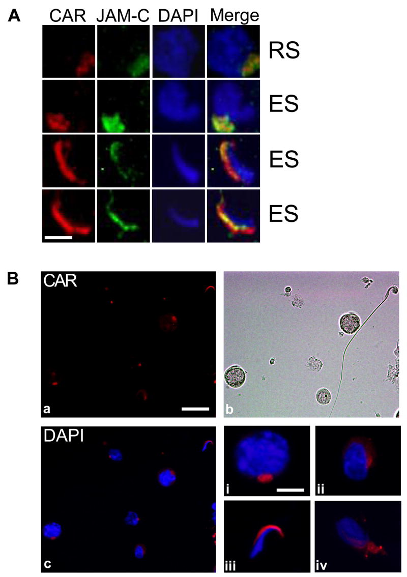
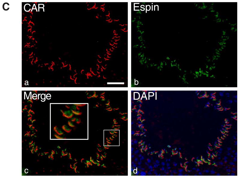
Fig. 6. CAR is expressed by germ cells at different stages of differentiation.
A: Immunofluorescent staining of rat testes sections shows co-localization of CAR and JAM-C in vivo. Both proteins were found to be distributed on round spermatids and were confined to the heads of elongating/elongate spermatids. Scale bar = 5 μm B: Rat germ cells were isolated by mechanical procedure (without glass wool), plated on poly-L-lysine coated coverslips and permeabilized before staining. (a) Localization of CAR on germ cells: Immunofluorescent staining was performed on the slides with rabbit anti-CAR (H-300) polyclonal antibody, followed by incubation with Cy3 conjugated donkey anti-rabbit IgG. Scale bar = 25 μm, which also applies to b & c. (b) Germ cells are visualized under light microscope. (c) Merged image of CAR and DAPI staining of nuclei. (i–iv) Magnified views of individual germ cells at different development stages. (i) spermatocyte (ii) round spermatid (iii) elongate spermatid (step17–18) (iv) elongating spermatid (step 9–10). Scale bar = 5 μm in (i), which applies to (i–iv). C: Immunofluorescent micrographs of a stage VIII seminiferous tubule. CAR (red) was stained with a rabbit anti-CAR (H-300) polyclonal antibody and espin (green) was stained with a mouse-anti espin monoclonal antibody. Merged image (c) shows that CAR and espin were colocalized to the apical ES site in the seminiferous epithelium. Scale bar= 50 μm.


