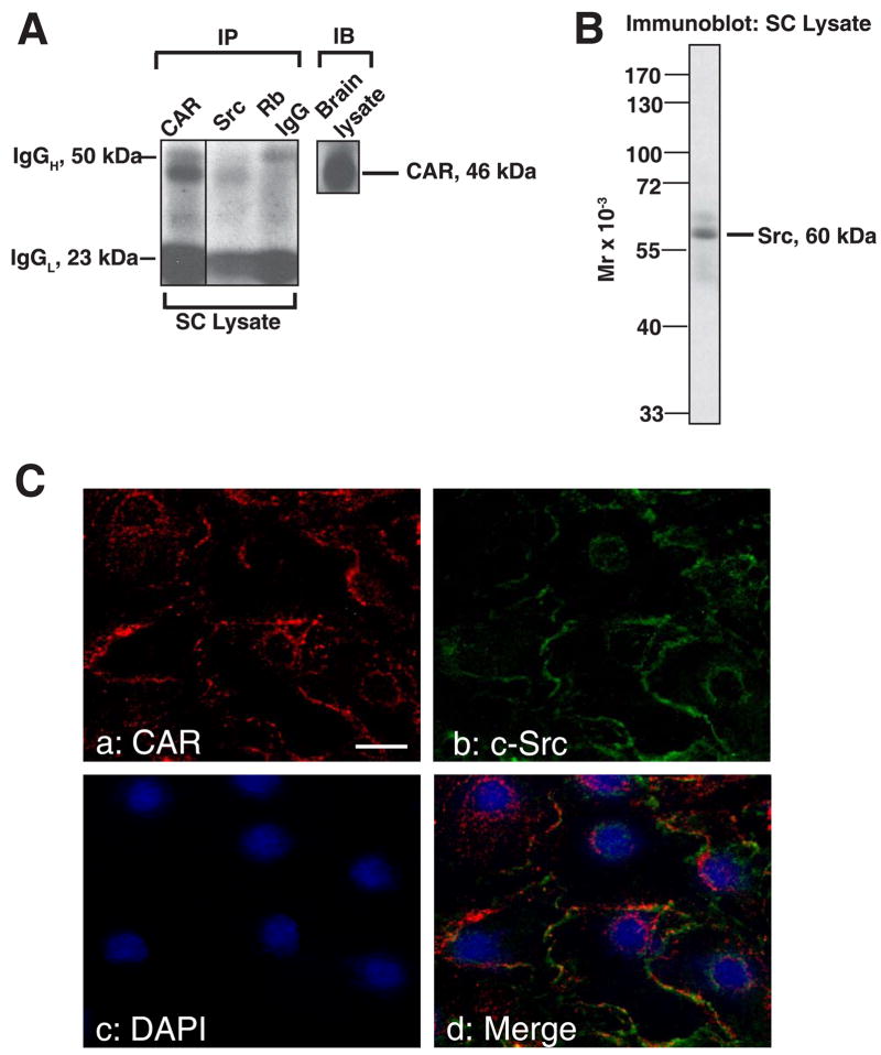
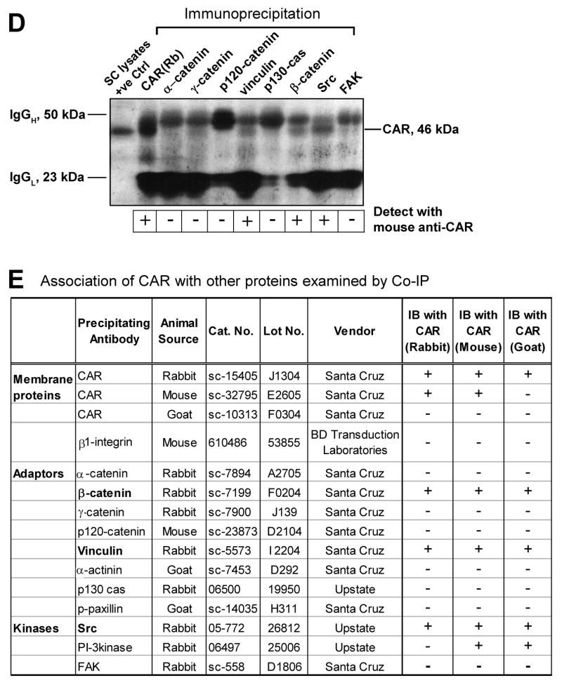
Fig. 8. Association between CAR and different adaptors and kinases in Sertoli cells cultured in vitro.
Sertoli cells were cultured for 4 days at 0.5 x 10⁶ cells/cm2 that permitted the establishment of functional tight and anchoring junctions, which also mimicked the functional physiology and morphology of Sertoli cells in vivo, prior to their use for lysate preparation. A: Co-IP experiments revealed the interaction between CAR and Src kinase in Sertoli cell lysates. 500 μg of Sertoli cell lysates were prepared and immunoprecipitated with rabbit monoclonal antibody towards Src kinase family. The immunocomplexes were then subject to immunoblot analysis and incubated with a mouse anti-CAR monoclonal antibody. Rabbit anti-CAR polyclonal antibody and normal rabbit IgG were also used as precipitating antibodies for the Co-IP experiments, serving as a positive and negative control, respectively. “Rb” stands for rabbit. B: A single prominent band corresponding to the apparent Mr of Src family protein kinase at 60 kDa was detected on the immunoblot using Sertoli cell lysate (100 μg protein), demonstrating the specificity of the antibody. C: Immunofluorescent staining reveals the colocalization of CAR with c-Src. Sertoli cells were cultured at low density (0.1 × 10⁶ cells/cm2) for 3 days before used for fluorescent microscopy. Cells were incubated with a rabbit anti-CAR polyclonal IgG and a mouse anti-c-Src monoclonal IgG as primary antibody, followed by a donkey-anti-rabbit CY3 (red fluorescence) conjugated secondary antibody (a) and a donkey-anti-mouse FITC (green fluorescence) (b) conjugated secondary antibody. Both proteins were seen to reside at cell-cell interface (a, b). Nuclei were visualized by DAPI staining (c). Areas of co-localization appear as orange (d). Scale bar = 30 μm in a, which applies to b-d. D-E: Aside from Src, we also performed co-IP studies with antibodies against other adaptors and protein kinase. In the blot shown in D, the immunocomplexes were subject to immunoblot analysis and incubated with a mouse anti-CAR monoclonal antibody. Lysates from Sertoli cells were loaded onto the same gel to illustrate the specificity of the antibody and served as positive control (+ve Ctrl). E: Tabulated co-IP results using different antibodies against protein adaptors and kinases present in Sertoli cells. Antibodies that failed to pull down CAR were also listed here. The immunocomplexes were separated by SDS-PAGE and probed with different CAR antibodies in immunoblots. The results shown here have been repeated three times using different batches of Sertoli cell cultures. “+”, positive co-IP result “−”, negative co-IP result.


