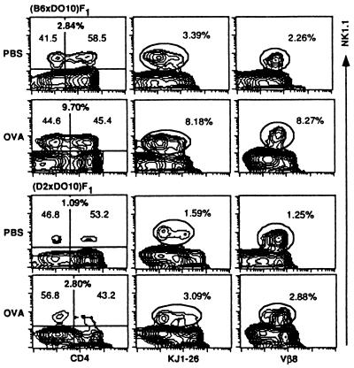
Figure 5.
Influence of cOVA administration on NK1.1+ thymocytes in (B6 × DO10)F1 and (D2 × DO10)F1 mice. Thymocytes from cOVA- or PBS-treated mice were isolated and stained with PE-anti-NK1.1, FITC-anti-CD4, and biotin-KJ1-26 or biotin-anti-Vβ8 followed by Streptavidin-FITC. A representative profile from three separate experiments is shown. The proportions of NK1.1+, NK1.1+ CD4−, and NK1.1+ CD4+ cells (Left), NK1.1+KJ1-26+ (NK1.1+KJ1-26low and NK1.1+KJ1-26intermediate) (Middle), and NK1.1+Vβ8+ (Right) cells are indicated.

