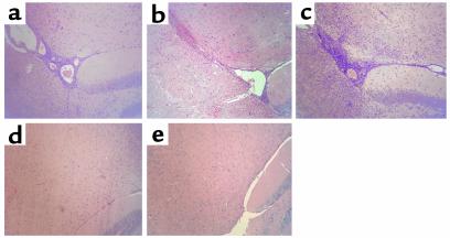
Figure 5.
Histological evidence of EAE in wild-type, Ii p31, and Ii p41, but not Ii- and H-2M–deficient, mice. Inflammatory lesions were observed in (a) wild-type, (b) Ii p31, and (c) Ii p41 mice. No evidence of inflammatory cell infiltration was seen in (d) Ii-deficient or (e) H-2M–deficient mice. CNS tissue was harvested, fixed, and stained with hematoxylin and eosin (H&E) as described in Methods. ×10.

