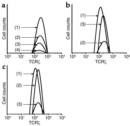
Figure 5.
Hp(2–20) (50 μM) triggers the disappearance of the signal-transducing molecule CD3ζ from NK cells. Monocytes and/or NK cell–enriched lymphocytes were incubated as described in the legend to Figure 4. After incubation of the lymphocyte/monocyte mixture, cells in a CD3ε– lymphocyte gate (>80% of which were CD56+ NK cells) were assayed for expression of CD3ζ by use of flow cytometry. The histogram in a shows CD3ζ expression by gated CD3ε– control lymphocytes + 25% monocytes (line 1), Hp(20–20)–treated lymphocytes + 25% monocytes (line 2), control lymphocytes + 50% monocytes (line 3), and Hp(2–20)–treated lymphocytes + 50% monocytes (line 4). The histogram in b shows corresponding CD3ζ expression of control CD3ε– lymphocytes (line 1), Hp(20–20)–treated lymphocytes + 25% monocytes (line 2), and Hp(20–20)–treated lymphocytes + 25% monocytes treated with catalase and SOD (line 3). The histogram in c shows CD3ζ expression of control CD3ε– lymphocytes (line 1), Hp(20–20)–treated lymphocytes + 25% monocytes + histamine (50 μM; line 2), and Hp(20–20)–treated lymphocytes + 25% monocytes + histamine + ranitidine (50 μM; line 3). The Hp(2–20) peptide was added 20 minutes after the onset of incubation. All data are expressed as percentage of CD3ζ+ cells in a CD3ε– gate, set to comprise only viable lymphocytes. Similar results were obtained in three separate experiments.

