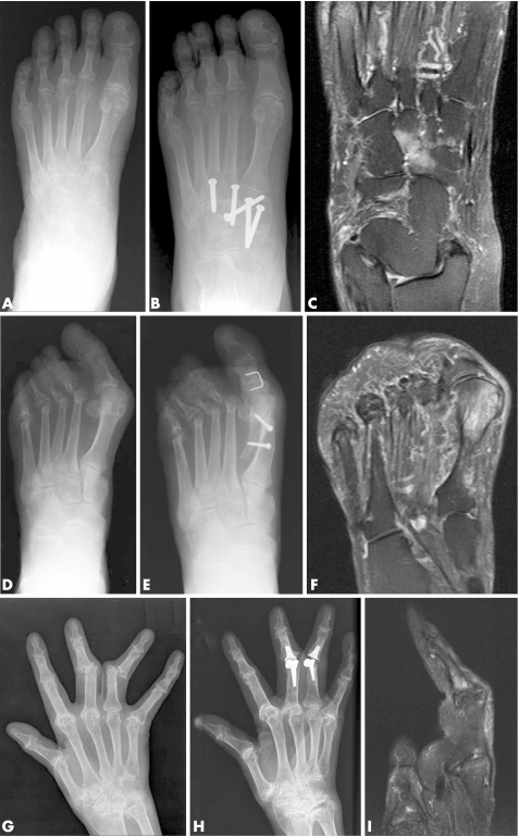
Figure 1 Bone oedema was seen frequently on the preoperative MRI scan within the field of intended surgery. (A) Patient 8. Preoperative radiograph of left foot showing reduced joint space at the tarsometatarsal joints and likely erosions “en face”. (B) Postoperative radiograph showing screws across naviculocuneiform and tarsometatarsal joints for arthrodesis. (C) Preoperative MRI scan of the foot (T2w coronal image) showing extensive bone oedema involving the medial, middle and lateral cuneiforms and navicular. (D) Patient 6. Preoperative radiograph of left foot showing severe hallux valgus and resected metatarsal heads (previous surgery). (E) Postoperative radiograph showing that a scarf osteotomy has been performed at the 1st metatarsal to improve alignment. (F) Preoperative MRI scan (T2w coronal image) showing extensive bone oedema involving the head of the 1st metatarsal extending to the mid‐shaft. (G) Patient 4. Preoperative radiograph of right hand showing advanced erosive damage of wrist, MCP and PIP joints with dislocation of 3rd and 4th PIP joints. (H) Postoperative radiograph showing joint replacements at 3rd and 4th PIP joints. (I) MRI scan of the 3rd finger of the right hand (sagittal T2w image) showing intense bone oedema involving the base of the middle phalanx and extending to the mid‐shaft.

An official website of the United States government
Here's how you know
Official websites use .gov
A
.gov website belongs to an official
government organization in the United States.
Secure .gov websites use HTTPS
A lock (
) or https:// means you've safely
connected to the .gov website. Share sensitive
information only on official, secure websites.
