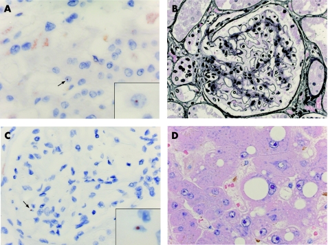
Figure 4 (A) Extensive steatosis in a liver specimen from a woman with systemic lupus erythematosus (SLE) (H&E staining). (B) Y‐chromosome‐positive cell (arrow, detail in inset) in the same liver as in (A). Red–brown dot indicates the Y‐chromosome, as identified by in situ hybridisation. (C) Lupus nephritis WHO class II in a renal specimen from a woman with SLE (silver staining). (D) Y‐chromosome‐positive cell (arrow, detail in inset) in the same kidney as in (C).

An official website of the United States government
Here's how you know
Official websites use .gov
A
.gov website belongs to an official
government organization in the United States.
Secure .gov websites use HTTPS
A lock (
) or https:// means you've safely
connected to the .gov website. Share sensitive
information only on official, secure websites.
