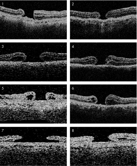
Figure 1 Pre‐operative optical coherence tomography of scans of patients 1–8 showing a “without cuff” macular hole configuration characterised by the absence of a distinct retinal cuff of fluid. The hole appears flat and punched out. The macular hole configuration of patient 7 is atypical. Although there is a cuff this hole rather fits into the “without cuff” category because the cuff is small, it does not overlap the hole, and it is not elevated.

An official website of the United States government
Here's how you know
Official websites use .gov
A
.gov website belongs to an official
government organization in the United States.
Secure .gov websites use HTTPS
A lock (
) or https:// means you've safely
connected to the .gov website. Share sensitive
information only on official, secure websites.
