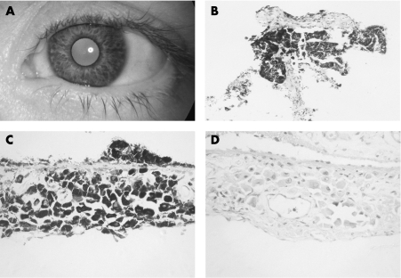
Figure 1 A: Anterior segment photograph of the left eye on presentation in 1995, showing dark, mainly flat patches of pigmentation. B: Initial biopsy specimen of inferonasal iris, showing a small iris fragment with large melanocytic cells on both sides of the iris (haematoxylin‐eosin ×200). C: Iridectomy specimen showing well‐orientated iris with large, densely pigmented, well‐separated cells involving the stroma (haematoxylin‐eosin ×400). D: Bleached section (×400) showing the morphology of the epithelioid cells in greater detail. Although most cells contain small nuclei, occasional cells show larger nuclei within which single nucleoli are present.

An official website of the United States government
Here's how you know
Official websites use .gov
A
.gov website belongs to an official
government organization in the United States.
Secure .gov websites use HTTPS
A lock (
) or https:// means you've safely
connected to the .gov website. Share sensitive
information only on official, secure websites.
