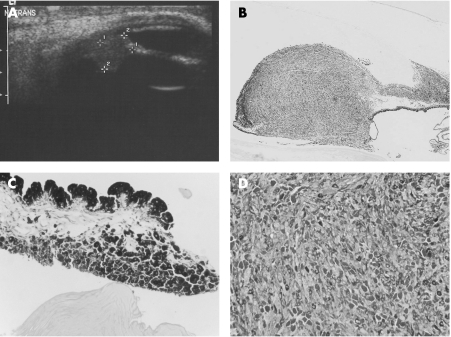
Figure 2 A: Anterior segment B mode ultrasound showing one of the two iris masses. This nasal tumour was elevated by 4.4 mm. B: The tumour is seen infiltrating the iris and iridocorneal angle of the enucleated globe under low power (haematoxylin‐eosin ×20). C: The iris has melanocytic cells on the posterior surface as well as a thickened sheet‐like proliferation on the anterior surface (haematoxylin‐eosin ×200). D: High‐power photomicrograph of the centre of the tumour, showing mixed epithelioid and spindle cell types (bleach ×200).

An official website of the United States government
Here's how you know
Official websites use .gov
A
.gov website belongs to an official
government organization in the United States.
Secure .gov websites use HTTPS
A lock (
) or https:// means you've safely
connected to the .gov website. Share sensitive
information only on official, secure websites.
