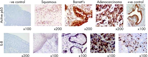
Figure 1 Example immunohistochemistry images with the active nuclear factor‐κB (NF‐κB) and interleukin‐8 (IL‐8) antibodies in squamous, Barrett's and adenocarcinoma sections, showing increasing staining across the histological series. Negative controls (no antibody) are shown for the active p65 antibody (adenocarcinoma material) and the IL‐8 antibody (Barrett's metaplasia material). Positive controls are also shown (breast ductal cancer for active p65 and tonsil for IL‐8).

An official website of the United States government
Here's how you know
Official websites use .gov
A
.gov website belongs to an official
government organization in the United States.
Secure .gov websites use HTTPS
A lock (
) or https:// means you've safely
connected to the .gov website. Share sensitive
information only on official, secure websites.
